Hip Socket
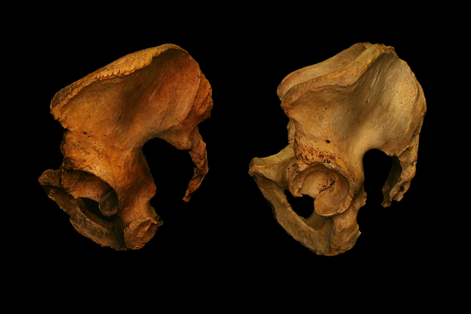
Left hip socket of two pelvises. The socket of the left specimen is pointed forward and down. The socket of the right specimen pointed sideways and nearly horizontal.

Hip sockets pointing in different directions. Therefore the range of motion in different directions will be different.

Hip sockets on right hand specimen are not visible from this front view because they are oriented out to the sides rather than forward.
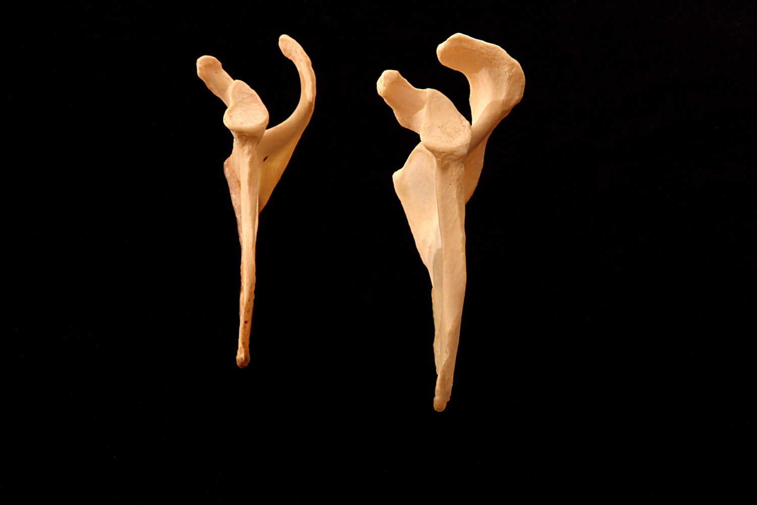
Femur

Six left femurs. Oriented upside down in this image to make it easier to show torsion in the next two images.

Looking down on the neck/head of six left femurs. The degree of torsion (twist) increases from left to right.

Two extremes of femoral torsion. Looking down on the neck/head of two left femurs. Feet parallel requires completely different femoral rotations.

Two left Femurs. The inclination of the neck is 40° different. The ability to abduct would be 40° different.

Two left femurs in front view. Specimen on the left has a neck that is twice as long.
Tibia

Two right tibia. The specimen on the left has external torsion. "Feet Parallel" would require an internal rotation of the tibia or femur.
Humerus

Looking down two right humerus bones. Specimen on left is torsioned internally. Therefore the humerus would have to externally rotate to place "hands parallel".
Scapula

Two right scapula from the back view. The acromion process on the left specimen doesn't cover the shoulder socket. Humerus abduction would vary tremendously.

Two right scapula. Left specimen could easily clasp hands behind the back. The acromion of the right specimen would block this movement.

Three right shoulder sockets of the scapula. The vertical axis of each is different. The acromion processes are also different.

Ventral view of two right scapula. Coracoid process on the right specimen is slanted downward, more likely to pinch with humerus in chaturanga.
Two left scapula. Acromion on left specimen doesn't cover shoulder socket, making it easy to raise arms past vertical.
Lumbar Vertebrae

Lumbar vertebrae. The gaps between spinous processes are different, so their back bending extensions would be different.
Sacrum

Three sacrum. Originally five bones fused in the womb. Different ratios of length to width due to the shape of bones.

Two sacrum. Originally five bones that fuse in the womb. Difference in curve due to difference in the five bones.